Allow me to introduce the "microtubule" to this forum.
This remarkable biochemical dipolar variable information processor.
Microtubules: the basics

Note that the "capping proteins" in the illustration are the synapses that connect neurons in neural networks
The reason why I believe microtubules are instrumental in the emergence of consciousness in living organisms is;
a) they are a dynamic information processor and a common denominator in all Eukaryotic organisms, and even earlier in a simpler form in Prokaryotic organisms.
b) they are the only known candidate meeting the requirements necessary for emergent levels of intelligence and consciousness.
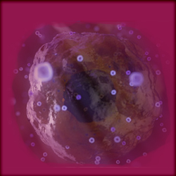
I believe that intelligence and emergent consciousness are observable at all levels of living biology, from motile single-celled organisms like the Paramecium and Slime Mold to heliotropism and photosynthesis in flora, to hive-intelligence, to self-aware consciousness in high order mammals.
IMO, there is but one candidate that meets all required properties that provide a functional network for the task of high-level information processing and that is the remarkably versatile microtubule.
A persuasive argument can be made on the presence of + 100 billion neural microtubules in the human brain connected by +100 trillion synapses.
There simply is nothing that can compare other than some magical concept. But as Tegmark observes, the brain contains all the required properties for conscious intelligence.
I believe this is the reason why Stuart Hameroff and (nobel laureate) Roger Penrose have proposed a theory of Orchestrated Objective Reduction (ORCH OR) that utilizes microtubules at a quantum level .
This remarkable biochemical dipolar variable information processor.
Microtubules: the basics
Microtubules: the basics
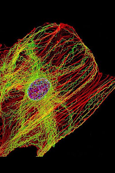
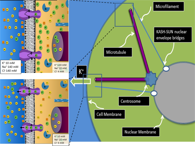
Microtubules are major components of the cytoskeleton. They are found in all eukaryotic cells, and they are involved in mitosis, cell motility, intracellular transport, and maintenance of cell shape.
Microtubules are composed of alpha- and beta-tubulin subunits assembled into linear protofilaments. A single microtubule contains 10 to 15 protofilaments (13 in mammalian cells) that wind together to form a 24 nm wide hollow cylinder.
https://www.nature.com/scitable/content/microtubules-the-basics-14673338/Microtubules are structures that can rapidly grow (via polymerization) or shrink (via depolymerization) in size, depending on how many tubulin molecules they contain.
Note that the "capping proteins" in the illustration are the synapses that connect neurons in neural networks
The reason why I believe microtubules are instrumental in the emergence of consciousness in living organisms is;
a) they are a dynamic information processor and a common denominator in all Eukaryotic organisms, and even earlier in a simpler form in Prokaryotic organisms.
b) they are the only known candidate meeting the requirements necessary for emergent levels of intelligence and consciousness.
I believe that intelligence and emergent consciousness are observable at all levels of living biology, from motile single-celled organisms like the Paramecium and Slime Mold to heliotropism and photosynthesis in flora, to hive-intelligence, to self-aware consciousness in high order mammals.
IMO, there is but one candidate that meets all required properties that provide a functional network for the task of high-level information processing and that is the remarkably versatile microtubule.
A persuasive argument can be made on the presence of + 100 billion neural microtubules in the human brain connected by +100 trillion synapses.
There simply is nothing that can compare other than some magical concept. But as Tegmark observes, the brain contains all the required properties for conscious intelligence.
I believe this is the reason why Stuart Hameroff and (nobel laureate) Roger Penrose have proposed a theory of Orchestrated Objective Reduction (ORCH OR) that utilizes microtubules at a quantum level .
Last edited: